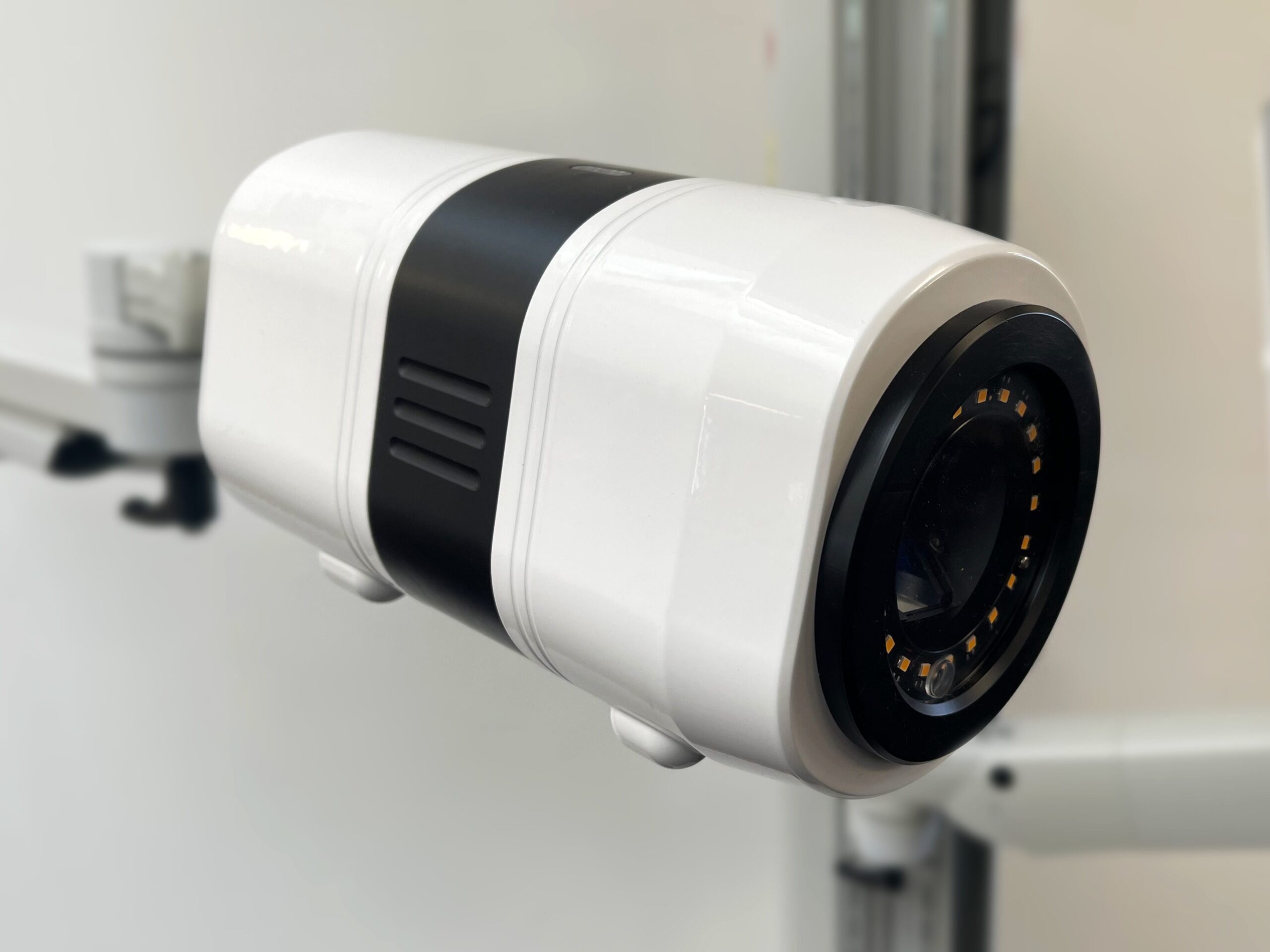
A high resolution hybrid gamma-optical camera which is versatile, compact and intuitive, designed to offer highly cost effective option for molecular imaging
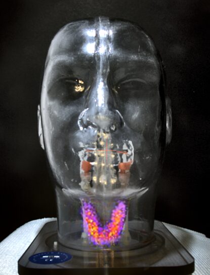
Fused, real-time, gamma-optical overlay
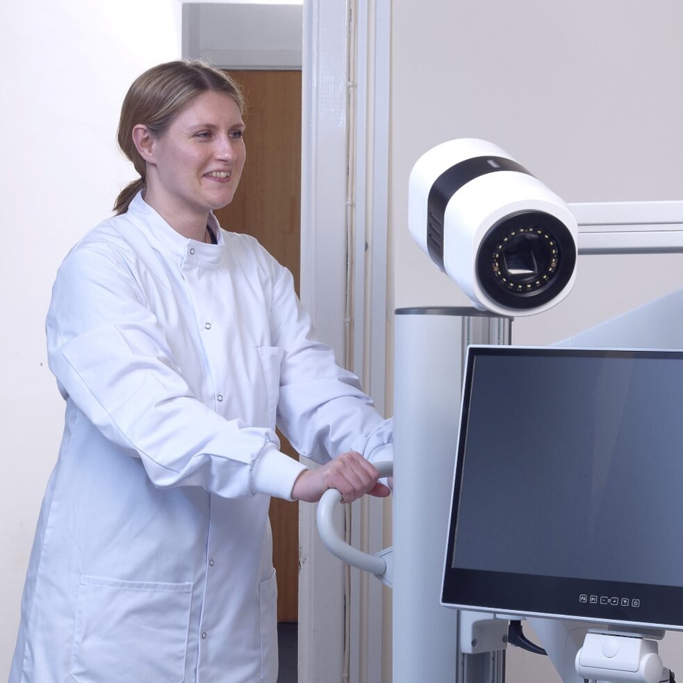
Operator-friendly imaging system: compact and easy to move
All this is packaged into a self-shielded camera head of 6 inch / 15cm diameter weighing ~5kg (about 10 pounds), mounted on a fully articulated arm for easy positioning and a wheel base that allows a single operator to transfer the system along corridors, through doorways and in elevators to any location within a hospital. A single cable connection enables plug-and-play operation for rapid installation and set up.
Seracam® is suitable for use with all commonly used isotopes for gamma imaging such as 99mTc, 123I, 111In (energy range 50-250 keV).
A touchscreen interface provides fast and intuitive camera control and real time image display for immediate operator feedback. The dedicated Seracam® application is designed for integration with hospital systems, with images provided in industry standard DICOM format.
Point-of-care imaging
Seracam® is truly portable and can be taken to any location within a hospital by a single operator. Plug-and-play connection means it is ready to use in any location within minutes.
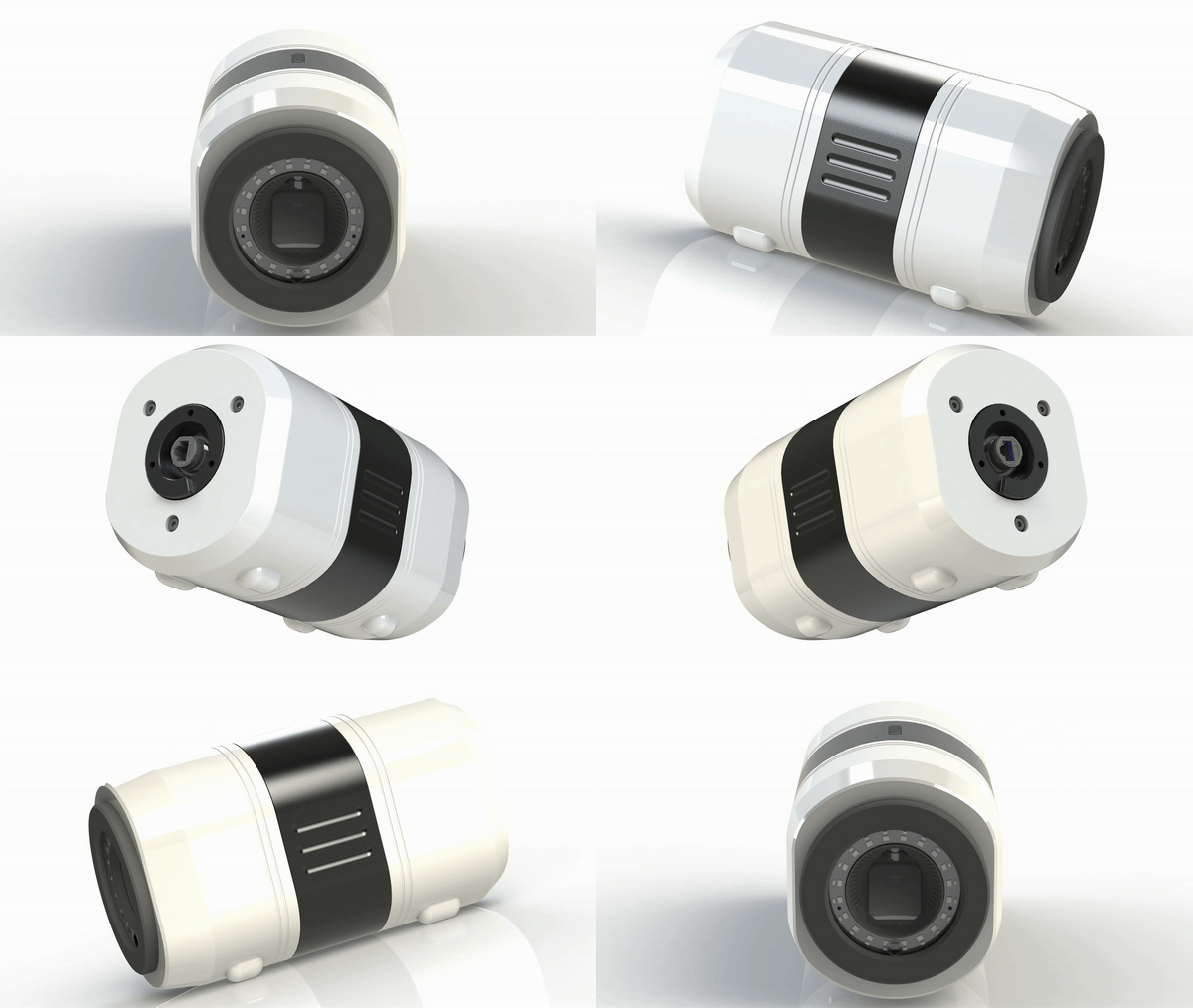
The small size means the camera can be placed closer to the patient than conventional large detectors to improve image resolution/acquisition times.
A selection of pinhole collimators allows the operator to determine the optimum balance of image resolution, acquisition speed and field of view for the case at hand.
The automatic change of aperture size provides a fast, flexible solution for many clinical applications without the operator dedicating a significant amount of time to change the collimator manually.
By providing an alternative to conventional gamma / SPECT cameras for imaging organs and structures within a small, portable footprint, Seracam® can both deliver further capacity within a nuclear medicine department without requiring an additional, dedicated camera room and provide access to molecular imaging at the point of care anywhere within the hospital.
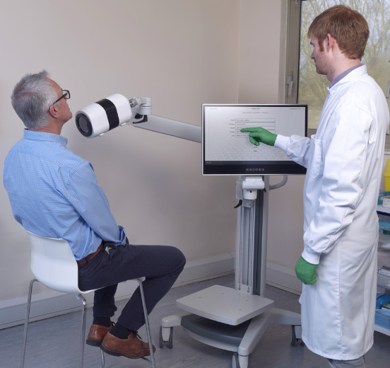
Improving the patient experience
Imaging at the patient’s bedside eliminates the need to bring the patient and nursing staff to the nuclear medicine department, which can be difficult for some patients (e.g. ICU, paediatric department). A fully articulated arm makes it easy to position the camera head for imaging in whatever position is most comfortable for the patient (e.g. sitting in a chair or wheelchair, or lying down on a bed or gurney).
Automatically co-registered gamma and optical images with fully aligned field-of-view and real time streaming of image display facilitates positioning for imaging, and the added visual information on uptake in relation to surface anatomy can improve communication on investigational findings with the patient and referring physician.
The small camera head presents a less intimidating experience for the patient than conventional full-body SPECT cameras, making it ideal for claustrophobic and paediatric patients.
A more comfortable patient is less prone to movement during the exam.
Transforming workflow in the Nuclear Medicine department
Current molecular imaging uses a mis-match of complex and expensive 3D imaging technologies for simple, small organ 2D imaging procedures.
Seracam can be used to free up capacity on these large fixed SPECT/CT imaging systems by providing an alternative solution for routine, simple, often time-consuming procedures (e.g. thyroid, gastric emptying, gall bladder).
