Medical Imaging
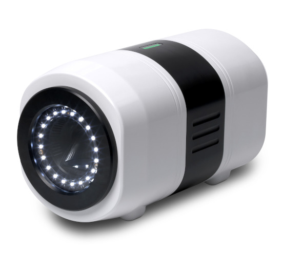
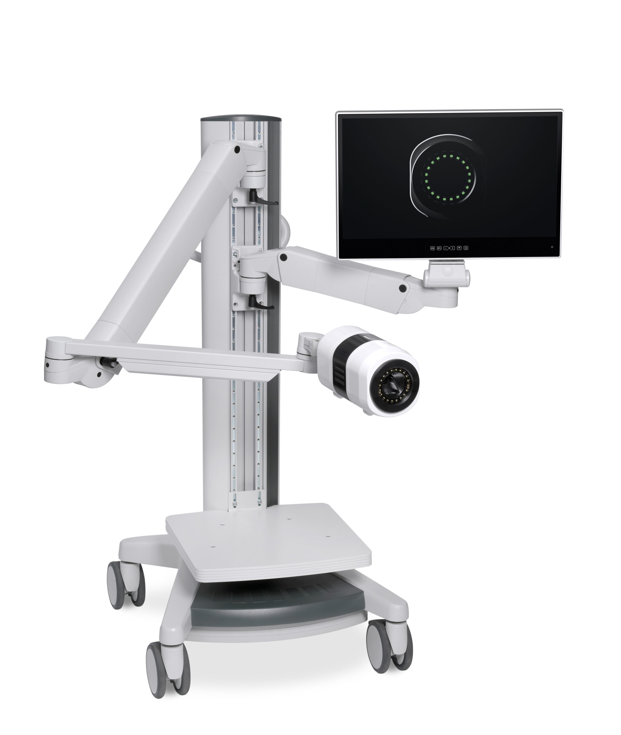
Seracam® is a game-changing molecular imaging technology designed to improve and increase patient access to care whilst reducing healthcare system costs
Currently the benefits of nuclear imaging, which uses gamma cameras to image the distribution of radiolabelled tracers within the body, are largely restricted to patients who can be referred to the nuclear medicine (NM) department of a hospital where the big, expensive, whole-body cameras are sited in dedicated rooms.
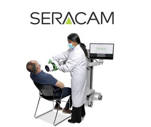
Seracam is designed to allow imaging to be routinely taken from the nuclear medicine department to the patient, wherever they may be – the outpatient clinic, hospital ward, physician’s office, the intensive care unit, or operating room.

Molecular Imaging and Personalised Medicine
Molecular imaging (MI) is a type of medical imaging that provides unique insights into what is happening inside the body at the cellular and molecular level helping physicians to deliver “personalised medicine” – i.e. delivering the right treatment to the right patient at the right time, leading to better outcomes, improved quality of life and reduced costs.
Unlike other medical imaging technologies such as x-rays, computed tomography (CT), magnetic resonance (MR) and ultrasound (US) which provide structural images, MI allows physicians to see how cells, tissues and organs are functioning and to measure chemical and biological processes without having to resort to biopsy or surgery.
MI procedures provide important information that help drive treatment decisions in patients with a range of conditions including cancer, infections, and disorders of the gastrointestinal, musculoskeletal and endocrine systems, kidney, heart, lung, and other organs.
Nuclear medicine (NM) is a form of MI in which images of the uptake of swallowed, inhaled or injested targeted radioactively-labelled tracers (radiopharmaceuticals) are captured with a gamma or SPECT camera.
For more information
Society of Nuclear Medicine and Molecular Imaging: About Nuclear Medicine and Molecular Imaging.
Standard Nuclear Medicine Procedures
- There are about 35+ different types of standard procedure conducted in nuclear medicine today, and a growing number of new procedures related to new therapies
- Many procedures do not gain any diagnostic benefit from increasingly complex technology yet standard of care dictates that both simple and complex procedures are performed with the same imaging techniques
- Specialised equipment like Seracam allows procedures to be conducted more efficiently by ensuring the correct procedure assignment to the most appropriate camera
- Workload efficiency improves making it possible to increase the number of patients scanned

Complex 3D &Simple/2D
Thyroid
Brain
Parathyroid
Bone
Sentinel Node
Lung
Gastric Emptying
Neuroendocrine
Gallbiadder
Simple/2D
Parathyroid
Sentinel Node
Gastric Emptying
Gallbiadder
Complex 3D
Brain
Bone
Lung
Neuroendocrine
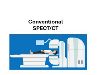
Currently large SPECT/CT scanners are used for all procedures – both complex and simple – exclusively in the nuclear medicine depertment
Seracam offers simple, small organ imaging at the point of care, for a patient centred experience.
Expensive SPECT cameras can be reserved for complex cases, leading to cost efficiency and improved workflow
Complex 3D & Simple/2D
Thyroid
Brain
Parathyroid
Bone
Sentinel Node
Lung
Gastric Emptying
Neuroendocrine
Gallbiadder
Simple/2D
Parathyroid
Sentinel Node
Gastric Emptying
Gallbiadder
Complex 3D
Brain
Bone
Lung
Neuroendocrine
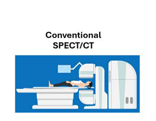
Currently large SPECT/CT scanners are used for all procedures – both complex and simple – exclusively in the nuclear medicine depertment
Expensive SPECT cameras can be reserved for complex cases, leading to cost efficiency and improved workflow
Nuclear Medicine Market Dynamics
- Incumbent manufacturers of traditional fixed SPECT/ SPECT-CT gamma cameras are creating increasingly sophisticated highly-featured systems with complex hardware
- Traditional fixed cameras are often the only systems available, even for relatively simple procedures
- There is an unmet medical need for high quality scans for simple procedures, improving speed and access for patients and transforming workflow in nuclear medicine departments.
Seracam®: platform technology
Seracam has been designed as a platform technology that can be easily modified to increase its clinical utility. For example, preliminary studies have shown that the optical camera can be readily modified to allow imaging of fluorescent markers, a capability that would be of particular value in surgical oncology. Applications outside the medical imaging field are also being investigated, such as nuclear operations.
