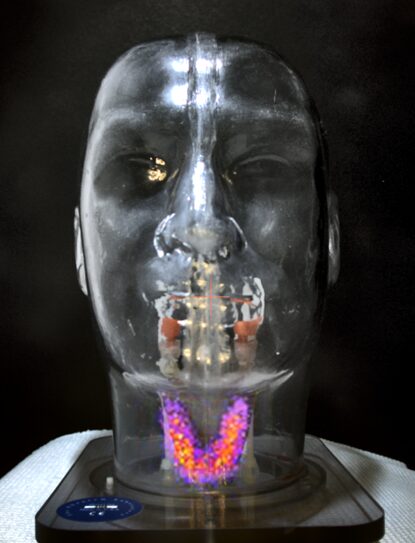
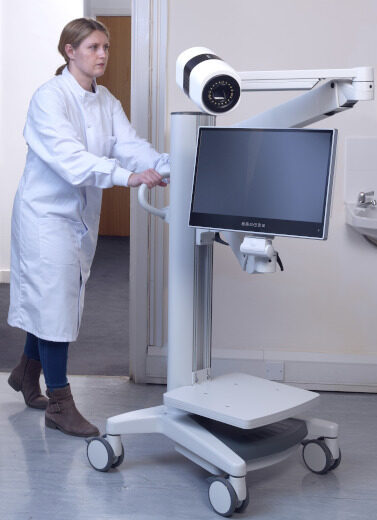
Serac Imaging Systems has developed Seracam®, a revolutionary and exceptionally versatile high resolution gamma-optical camera, aiming to bring nuclear imaging to the patient bedside
Seracam leverages technology designed for NASA space observatories to match or exceed the performance of the current class of systems with greater ease of use for small organ imaging procedures. Seracam has been designed from the outset for its practical application, with its compact design and intuitive operating system enabling imaging at the point of care. This will ease pressure on complex SPECT/ CT cameras by providing an efficient and cost-effective alternative for conducting many routine, simple, but often time-consuming procedures.
This technology has the potential to improve patient access to care, improve workflow in the nuclear medicine department, and reduce healthcare system costs.
Seracam is currently being tested at a number of clinical sites ahead of regulatory submission.
For information on industrial applications for Seracam please see here.
Unique Hybrid gamma-optical camera
The automatic overlay of the gamma and optical images, with matched fields of view and alignment maintained at any imaging angle or distance, gives clinicians precision imaging.
Such enhanced imaging technology has the potential to help clinicians make better, more informed and more timely treatment decisions. The optical overlay also enhances patient understanding of the images.

Point-of-Care Imaging
Seracam is designed to allow imaging to be routinely taken from the nuclear medicine department to the patient, wherever they may be – the outpatient clinic, hospital ward, physician’s office, the intensive care unit, or operating theatre.
Currently, this imaging is largely restricted to patients who can be referred to the nuclear medicine department of a hospital where large, expensive, heavy and complex cameras are sited in dedicated rooms.

Types of standard nuclear medicine procedures are routinely performed in a nuclear medicine department
0
+
Nuclear medicine facilities globally
0
Nuclear medicine procedures conducted per annum
0
m
Seracam® Features
Simple and intuitive
Unique hybrid gamma-optical imaging enhances utility and usability
Highly versatile, easily relocated for point-of-care imaging
Small and patient friendly
Flexible head for multiple scanning applications
Affordable
Same-day installation, minimal service requirements and no downtime

Our Technology
Seracam®: Game-changing gamma imaging made easy
Like all gamma cameras, Seracam is used to form images revealing the distribution of a radiopharmaceutical that has been administered to a patient in order to help in the diagnosis or monitoring of disease.
Seracam® uses a microcolumnar CsI(Tl) crystal scintillator to convert gamma photons to optical photons that are detected by a semi-conductor.

Fresh and Novel Design Approach
- Designed from the outset for ease of use, compact design, and truly mobile for imaging at the point-of-care
- Unconstrained by pre-conceived ideas from traditional nuclear medicine imaging systems
- Incorporates cutting-edge technologies from other industries including technology designed for NASA space observatories to deliver equal or better performance than the current standard of care
Investment Opportunities
Serac Imaging Systems is a privately owned, clinical stage medical imaging company. We are always ready to talk to investors who match our ambitions and can help us excel. If you would like to discuss investment opportunities, please do get in touch.

Meet the Team
Discover more about the team behind Serac Imaging Systems.
Latest News
05/11/2025
New Seracam® Extravasation Imaging Application and Enhanced Intraoperative Capabilities Presented at IEEE
Serac Imaging Systems announces that two posters featuring Seracam® are being presented by its academic partners from Loughborough University at the IEEE Nuclear Science Symposium, Medical Imaging Conference, taking place from 1-8 November 2025 in Yokohama, Japan. One displays the first presentation of Seracam®’s potential to prevent extravasation injury; the other introduces new features to enhance Seracam®’s capabilities in intraoperative imaging.
01/10/2025
Small Organ and Intraoperative Imaging with Seracam® Presented at IUPESM
Serac Imaging Systems and its clinical investigators from the Faculty of Medicine at the University of Malaya, Malaysia, announce that an oral presentation evaluating the first clinical experience using Seracam® took place at the IUPESM World Congress on Medical Physics and Biomedical Engineering in Adelaide, Australia (29 September to 4 October).
25/06/2025
Engineering Magazine
Seracam® features in the June 2025 edition of Engineering Magazine.

Seracam® is for investigational use only and has not been registered or approved by the FDA, UK, European or Malaysian regulatory authorities.
